Have growers worried about Neopestalotiopsis on strawberries?
These NPDN labs can help identify aggressive strains
Ava Veith (1), Marcus Vinicius Marin (2), Alejandra Jimenez Madrid* (3), Xiao Yang (4), Francesca Rotondo (5), Lina Rodriguez Salamanca* (1), Natalia A. Peres (2)
(1) Virginia Tech, (2) University of Florida, Gulf Coast Research and Extension Center, (3) University of Georgia – Tifton, (4) Department of Plant Industry, Regulatory Services, Clemson University, (5) The Ohio State University – Wooster
*Members of the NPDN Protocol and Validation committee
The Communicator: Volume 6, Issue 4, April 2025
Neopestalotiopsis is an emerging pathogen that infects strawberry, causing leaf spot, fruit rot, crown rot, root rot, and lesions on petioles and stolons, all leading to yield loss. The disease is most severe under prolonged periods of wetness (>24h) and is a major concern for nurseries and fruit producers. Due to the presence of both aggressive and non-aggressive strains, early detection of aggressive strains is crucial for effective management. This article aims to provide new diagnostic information and resources for detecting Neopestalotiopsis on strawberry.
Symptoms and Signs
Neopestalotiopsis affects all plant parts, including fruits, leaves, crowns, and roots. Key symptoms include dry, circular or irregularly shaped lesions, light tan to brown in color, which become darker as they expand. Sunken lesions also can be observed on petioles and fruit, along with crown rot (which typically starts from the outer tissues and is characterized by orange or brown discoloration), as well as root decay (Figure 1). Black acervuli may develop on old necrotic foliar lesions and fruit.
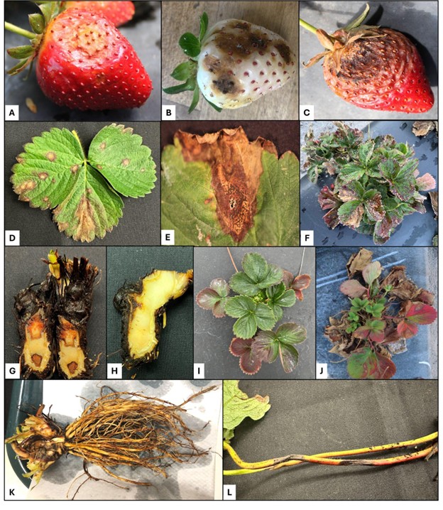
Figure 1. Signs and symptoms associated with Neopestalotiopsis diseases of strawberry. Early stage of fruit rot symptoms with sunken lesions on a red fruit (A) and a white fruit (B); C, late-stage symptoms on a fruit containing black acervuli; D, Light-brownish leaf spots; E, Necrotic tissue bearing acervuli and cirrhi; F, Top view of a plant severely affected with spots, necrosis, and blight; G, Crown rot with internal discoloration; H, Crown rot developed from the outer tissues; I,J Plants with stunted growth showing purplish to reddish leaves; K, Roots showing lesions and rot; L, Sunken lesions on petioles. (reprinted with permission from Marcus Marin, and Strawberry Pathology Lab - UF, GCREC)
Recommended Isolation Procedures
To isolate Neopestalotiopsis from symptomatic strawberry tissue, cut 5x5 mm pieces from the transition zone between healthy and necrotic tissue, disinfect in 0.25-0.3% sodium hypochlorite for 1 min, rinse with sterile water, and transfer to a general isolation medium (19.5g PDA, 9.5g agar, 0.1g streptomycin, 0.25g ampicillin per liter). Alternatively, isolate by transferring acervuli from sporulating lesions or plating disinfected crown, root, and petiole pieces. Adding 75 ppm chloramphenicol to PDA or using acidified PDA (APDA) also is effective (Jimenez Madrid et al., 2024). Incubate plates at 25°C for 5-6 days (Kaur et al., 2023). After approximately seven days of growth on PDA, white fungal colonies with circular, wavy edges and a cottony upper surface become visible, regardless of the isolate (Baggio et al., 2021).
How do we differentiate aggressive strains from saprophytic?
Neopestalotiopsis was first reported as a strawberry pathogen in 1973 (Howard and Albregts, 1973), but it has been considered a minor pathogen associated with stressed plants. Its taxonomy remains unclear, and the emergence of aggressive strains complicates disease diagnostics and management. Aggressive and non-aggressive strains are morphologically indistinguishable, so molecular techniques such as PCR/RFLP and high-resolution melting (HRM) assays developed based on a β-tubulin gene fragment are reliable methods for accurate diagnosis. The PCR/RFLP assay produces two bands after digestion for aggressive Neopestalotiopsis sp., while a single band can be found for N. rosae (Kaur et al., 2023). The HRM assay can distinguish aggressive Neopestalotiopsis sp. from N. rosae and other species based on different melting peaks (Rebello et al., 2023). Both markers work directly to amplify spores and aerial mycelium without prior DNA extraction. These molecular methods were designed to differentiate the aggressive Neopestalotiopsis sp. strain first detected in Florida in 2017, which spread across the U.S. (Rebello et al., 2023). However, a second aggressive strain, identified as N. rosae in Mexico in 2017, was introduced to the U.S. in 2022–2023 and may cause false negatives with the current methods. With the emergence of this new strain, isolates in the N. rosae group can now be either aggressive or non-aggressive. The second strain first reported from Mexico has been confirmed to be present in FL (Marin and Peres, personal communication), SC (Yang, personal communication), and Georgia (Jimenez Madrid, personal communication). To improve diagnostic tools, the UF-GCREC Plant Clinic has used whole-genome sequencing to develop new primers that detect only aggressive phenotypes from both strains via HRM assay. This method is unpublished, but diagnosticians, researchers or growers can send samples for testing to the diagnostic labs that offer the specialized testing.
Sample type
For accurate disease diagnosis, strawberry samples should be collected at early symptom development stages, with plant parts that remain partially alive and green. Completely necrotic, dried, or long-dead samples are unsuitable, as they often contain secondary microbes (including non-aggressive Neopestalotiopsis) that can limit the identification of the primary pathogen. Samples should be collected before pesticide application, kept refrigerated, and never exposed to heat or moisture before submission. A summary of the diagnostic workflow is presented in Figure 2.
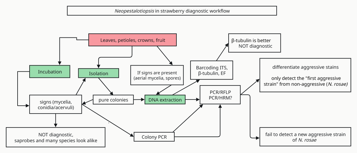
Figure 2. Flow chart describing different techniques in which Neopestalotiopsis infecting strawberry plants can be identified.
What NPDN labs can help?
Listed below are diagnostic labs which offer specialized testing for identifying pathogens in affected samples submitted by other diagnosticians, researchers, or growers.
University of Florida Gulf Coast Research and Education Center’s Plant Clinic Location: Wimauma, FL; SPDN
Fee: Free for FSGA members, non-members charged $40 per sample
Services:
- Standard culture isolation and HRM assays (includes new primers to detect both aggressive strains of Neopestalotiopsis
- Plant samples or Neopestalotiopsis cultures may be submitted and must be accompanied by a completed Strawberry Sample Submission form
Contact Information:
Dr. Natalia Peres – nperes@ufl.edu
Dr. Marcus Marin – marin.m@ufl.edu
813-419-6599
University of Georgia Plant Molecular Diagnostic Laboratory Location: Tifton, GA; SPDN
Fee: $60 in-state and out-of-state; additional $20 charge if DNA sequencing needs to be done to confirm identification.
Services:
- Fungal isolation, DNA extraction, PCR/RFLP assay, gel electrophoresis following the Kaur et al (2023) protocol
- Plant samples and Neopestalotiopsis cultures are accepted
- Sample collection, submission, and fee information available on the Plant Molecular Diagnostic Laboratory website
Contact Information:
Dr. Alejandra Jimenez Madrid – a.jimenez@uga.edu
229-386-3372
The Ohio State University C. Wayne Ellett Plant and Pest Diagnostic Clinic Location: Wooster, OH; NCPDN
Fee:
Free for Ohio fruit and vegetable open-field growers (supported by the Ohio Small Fruit and Vegetable Development Program 2025)
$30 for remaining in-state stakeholders
$50 out-of-state
Services:
- Culturing and molecular characterization based on PCR/RFLP assay, gel electrophoresis following the Kaur et al (2023) protocol
- Submit samples to:
Selby Hall
1680 Madison Avenue
Wooster, OH 44691
Contact Information:
Dr. Francesca Rotondo – rotondo.11@osu.edu
330-263-3721
More information on lab website
Clemson University Plant and Pest Diagnostic Clinic Location: Pendleton, SC; SPDN
Fees and Services:
- Isolation and incubation ($20 in-state / $30 out-of-state)
- PCR-RFLP (Free for SC clients / $50 out-of-state)F
Contact Information:
Dr. Xiao Yang – xyang7@clemson.edu
864-646-2133
Ongoing DRAFT diagnostic guide in progress Neopestalotiopsis in Strawberry (view only)
References
Baggio, J. S., Forcelini, B. B., Wang, N.-Y., Ruschel, R. G., Mertely, J. C., & Peres, N. A. (2021). Outbreak of leaf spot and fruit rot in florida strawberry caused by Neopestalotiopsis spp. Plant Disease, 105(2), Article 2. https://doi.org/10.1094/PDIS-06-20-1290-RE
Kaur, H., Gelain, J., Marin, M. V., Peres, N. A., & Schnabel, G. (2023). Development of a molecular tool for identification of a new Neopestalotiopsis sp. associated with disease outbreaks on strawberry. Plant Disease, 107(5), Article 5. https://doi.org/10.1094/PDIS-09-22-2117-RE
McNally, J., Prapagar, K., Goldenhar, K., Pate, E., Shan, S., & Kalischuk, M. (2023). First report of an aggressive species of Neopestalotiopsis affecting strawberry in Canada. New Disease Reports, 48(1), Article 1. https://doi.org/10.1002/ndr2.12210
Han, H., Jang, Y. J., Oh, Y., Marin, M. V., Huguet-Tapia, J., Peres, N. A., & Lee, S. (2024). Chromosome-scale genome sequence resource for two Neopestalotiopsis spp. isolates with different virulence in strawberry (Fragaria × ananassa). PhytoFrontiersTM, 4(3), 422–426. https://doi.org/10.1094/PHYTOFR-08-23-0110-A
Howard, C.M. and E.E. Albregts (1973). A strawberry fruit rot caused by Pestalotia longisetula. Phytopathology, 63:862-863.
Jimenez Madrid, A., Munoz, G., Collins, C., & Brannen, P. (2024). First report of the new Neopestalotiopsis species causing strawberry leaf spot and fruit rot in Georgia. Plant Disease, 108(8), 2574. https://doi.org/10.1094/PDIS-02-24-0409-PDN
Jimenez Madrid, A. Neopestalotiopsis Leaf Spot and Fruit Rot Disease | Southern Region Small Fruit Consortium. (2025, January 21). https://smallfruits.org/2025/01/survey-of-neopestalotiopsis-leaf-spot-and-fruit-rot-disease-in-the-southeastern-us/
Rebello, C. S., Wang, N.-Y., Marin, M. V., Baggio, J. S., & Peres, N. A. (2023). Detection and species differentiation of Neopestalotiopsis spp. from strawberry (Fragaria × ananassa) in Florida using a high-resolution melting analysis. PhytoFrontiersTM, 3(1), 156–163. https://doi.org/10.1094/PHYTOFR-03-22-0034-FI
Rebollar-Alviter, A., Silva-Rojas, H. V., Fuentes-Aragón, D., Acosta-González, U., Martínez-Ruiz, M., & Parra-Robles, B. E. (2020). An emerging strawberry fungal disease associated with root rot, crown rot and leaf spot caused by Neopestalotiopsis rosae in Mexico. Plant Disease, 104(8), 2054–2059. https://doi.org/10.1094/PDIS-11-19-2493-SC
Small Fruits (2023, October). Laboratories that offer testing. Southeastern US Small Fruit Consortium. https://smallfruits.org/2023/10/laboratories-that-offer-testing/#:~:text=Plant%20Molecular%20Diagnostics%20Lab&text=UGA's%20MDL%20also%20tests%20for,is%20an%20additional%20%2420%20charge